A Guide to Mastering Lower Extremity Manual Muscle Testing with Handheld Dynamometers
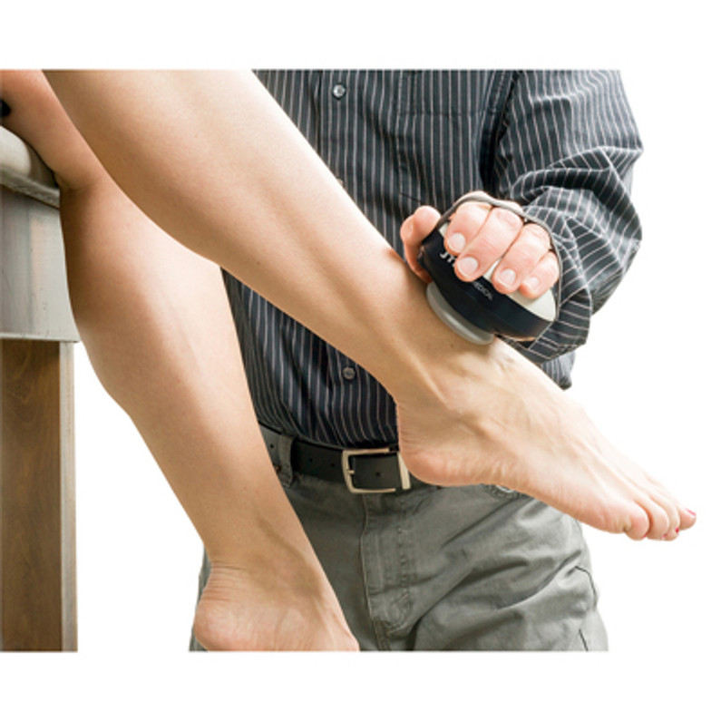
 Manual Muscle Testing (MMT) with handheld dynamometers is a fundamental component of musculoskeletal assessment in rehabilitation settings. These portable devices provide objective measurements of muscle strength that aid clinicians in evaluating and monitoring patients' progress accurately. In this article, we'll explore the specific techniques and instructions for performing manual muscle tests using handheld dynamometers for the body's lower extremities.
Hip Flexion
The hip flexion, the movement of lifting the thigh toward the abdomen, is a critical action involved in various daily activities, such as walking, climbing stairs, and rising from a seated position.
The primary muscle responsible for this movement is the iliopsoas, which consists of the iliacus and the psoas major. Additionally, other muscles, such as the rectus femoris, sartorius, and tensor fasciae latae, contribute to hip flexion to varying degrees.
Testing Procedure for Hip Flexion: Performing manual muscle testing for hip flexion involves several key steps to ensure accuracy and reliability.
- Positioning: Position the patient comfortably in a supine position on a treatment table, with both legs fully extended.
- Stabilization: Stabilize the pelvis to isolate the movement to the hip joint. You can achieve this by providing manual stabilization of the contralateral hip with your non-testing hand or by using a belt or strap to secure the pelvis.
- Palpation: Identify the insertion point of the iliopsoas, which is typically located on the lesser trochanter of the femur.
- Testing: Instruct the patient to lift their leg upward, bending at the hip joint and bringing the thigh toward the abdomen.
- Application of Resistance: Place one hand on the distal thigh or knee of the patient's testing leg to provide resistance. Apply pressure in the direction opposite to hip flexion, ensuring a gradual increase in resistance to assess the muscle's strength throughout the range of motion.
- Observation: Observe the patient's ability to maintain the position against resistance. Look for compensatory movements or substitutions, such as trunk flexion or pelvic tilting, which may indicate weakness in the targeted muscle or muscles.
- Grading: Use a standardized grading scale, such as the Medical Research Council (MRC) scale or the Modified Oxford Scale, to assign a muscle grade based on the patient's performance. Grades typically range from 0 to 5, with 0 indicating no muscle contraction and 5 representing normal strength.
Hip Extension
The hip extension, the movement of moving the thigh backward, is vital for various activities like walking, running, and climbing stairs.
The main muscle responsible for this movement is the gluteus maximus, the largest muscle in the body. Additionally, the hamstrings, particularly the biceps femoris, also play a significant role in hip extension.
Testing Procedure for Hip Extension: Performing manual muscle testing for hip extension involves several key steps to ensure accuracy and reliability.
- Positioning: Begin by positioning the patient prone on a treatment table, with the lower limbs relaxed and hanging off the edge.
- Stabilization: Stabilize the pelvis to isolate the movement to the hip joint. You can achieve this by firmly pressing down on the contralateral iliac crest or by having the patient hold onto the table for stability.
- Palpation: Identify the insertion point of the gluteus maximus, which typically lies on the posterior aspect of the femur and the iliotibial band.
- Testing: Instruct the patient to lift their leg off the table by extending the hip, attempting to bring the thigh in line with the body.
- Application of Resistance: Apply resistance against the patient's lower leg, just above the ankle, in a direction opposing hip extension. Gradually increase the resistance while ensuring that the patient can maintain the position against it.
- Observation: Observe the patient's ability to maintain the position and note any compensatory movements, such as lumbar hyperextension or contralateral hip abduction, which may indicate weakness or compensation.
- Grading: Utilize a standardized grading scale, such as the Medical Research Council (MRC) scale, to assign a muscle grade based on the patient's performance. This scale ranges from 0 to 5, with 0 indicating no muscle contraction and 5 representing normal strength.
Hip Abduction
The hip abduction, the movement of lifting the leg away from the body's midline, is essential for various activities like walking, running, and maintaining balance.
The primary muscle responsible for this movement is the gluteus medius, located on the lateral aspect of the hip. Additionally, the tensor fasciae latae (TFL) also contributes to hip abduction to a lesser extent.
Testing Procedure for Hip Abduction: Performing manual muscle testing for hip abduction involves several key steps to ensure accuracy and reliability.
- Positioning: Begin by positioning the patient lying on their side on a treatment table, with the lower leg slightly flexed at the knee for stability.
- Stabilization: Stabilize the pelvis to isolate the movement to the hip joint. Place one hand on the iliac crest of the uppermost hip to prevent pelvic rotation during the test.
- Palpation: Identify the muscle belly of the gluteus medius, which lies beneath the iliac crest on the lateral aspect of the hip.
- Testing: Instruct the patient to lift the top leg upward, away from the body's midline, while keeping the knee straight.
- Application of Resistance: Apply resistance against the patient's leg, just above the knee, in a direction opposing hip abduction. Gradually increase the resistance while ensuring that the patient can maintain the position against it.
- Observation: Observe the patient's ability to maintain the position and note any compensatory movements, such as pelvic rotation or trunk lateral flexion, which may indicate weakness or compensation.
- Grading: Utilize a standardized grading scale, such as the Medical Research Council (MRC) scale, to assign a muscle grade based on the patient's performance. This scale ranges from 0 to 5, with 0 indicating no muscle contraction and 5 representing normal strength.
Hip Adduction
The hip adduction, the movement of bringing the leg toward the body's midline, is crucial for various activities such as walking, maintaining balance, and certain athletic movements.
The main muscle responsible for this movement is the adductor magnus, located on the inner thigh. Additionally, the adductor longus, adductor brevis, and gracilis also contribute significantly to hip adduction.
Testing Procedure: Performing manual muscle testing for hip adduction involves several key steps to ensure accuracy and reliability.
- Positioning: Begin by positioning the patient lying on their side on a treatment table, with the lower leg slightly flexed at the knee for stability.
- Stabilization: Stabilize the pelvis to isolate the movement to the hip joint. Place one hand on the iliac crest of the uppermost hip to prevent pelvic rotation during the test.
- Palpation: Identify the muscle bellies of the adductor muscles, which lie along the inner thigh.
- Testing: Instruct the patient to lift the bottom leg upward, toward the body's midline, while keeping the knee straight.
- Application of Resistance: Apply resistance against the patient's leg, just above the knee, in a direction opposing hip adduction. Gradually increase the resistance while ensuring that the patient can maintain the position against it.
- Observation: Observe the patient's ability to maintain the position and note any compensatory movements, such as pelvic rotation or trunk lateral flexion, which may indicate weakness or compensation.
- Grading: Utilize a standardized grading scale, such as the Medical Research Council (MRC) scale, to assign a muscle grade based on the patient's performance. This scale ranges from 0 to 5, with 0 indicating no muscle contraction and 5 representing normal strength.
Hip External Rotation
The hip external rotation, the movement of rotating the thigh away from the body's midline, is essential for various activities such as walking, running, and maintaining balance.
The primary muscles responsible for this movement are the deep external rotators of the hip, including the piriformis, gemellus superior, gemellus inferior, obturator internus, and quadratus femoris. Additionally, the gluteus maximus and gluteus medius also contribute to hip external rotation to varying degrees.
Testing Procedure Hip External Rotation: Performing manual muscle testing for hip external rotation involves several key steps to ensure accuracy and reliability.
- Positioning: Begin by positioning the patient lying on their side on a treatment table, with the lower leg slightly flexed at the knee for stability. Ensure that the pelvis is aligned with the edge of the table.
- Stabilization: Stabilize the pelvis to isolate the movement to the hip joint. Place one hand on the contralateral hip to prevent pelvic rotation during the test.
- Palpation: Identify the muscle bellies of the deep external rotators, which are located on the lateral aspect of the hip joint.
- Testing: Instruct the patient to lift the top leg upward, away from the body's midline, while keeping the knee bent at a 90-degree angle.
- Application of Resistance: Apply resistance against the patient's leg, just above the ankle, in a direction opposing hip external rotation. Gradually increase the resistance while ensuring that the patient can maintain the position against it.
- Observation: Observe the patient's ability to maintain the position and note any compensatory movements, such as pelvic rotation or trunk lateral flexion, which may indicate weakness or compensation.
- Grading: Utilize a standardized grading scale, such as the Medical Research Council (MRC) scale, to assign a muscle grade based on the patient's performance. This scale ranges from 0 to 5, with 0 indicating no muscle contraction and 5 representing normal strength.
Hip Internal Rotation
Hip internal rotation, the movement of rotating the thigh toward the body's midline, is crucial for various activities such as walking, running, and maintaining balance.
The primary muscles responsible for this movement are the hip internal rotators, including the gluteus minimus, tensor fasciae latae (TFL), anterior fibers of the gluteus medius, and the anterior fibers of the adductor magnus.
Testing Procedure Hip Internal Rotation: Performing manual muscle testing for hip internal rotation involves several key steps to ensure accuracy and reliability.
- Positioning: Begin by positioning the patient lying on their side on a treatment table, with the lower leg slightly flexed at the knee for stability. Ensure that the pelvis is aligned with the edge of the table.
- Stabilization: Stabilize the pelvis to isolate the movement to the hip joint. Place one hand on the contralateral hip to prevent pelvic rotation during the test.
- Palpation: Identify the muscle bellies of the hip internal rotators, which are located on the inner aspect of the hip joint.
- Testing: Instruct the patient to lift the bottom leg upward, toward the body's midline, while keeping the knee bent at a 90-degree angle.
- Application of Resistance: Apply resistance against the patient's leg, just above the ankle, in a direction opposing hip internal rotation. Gradually increase the resistance while ensuring that the patient can maintain the position against it.
- Observation: Observe the patient's ability to maintain the position and note any compensatory movements, such as pelvic rotation or trunk lateral flexion, which may indicate weakness or compensation.
- Grading: Utilize a standardized grading scale, such as the Medical Research Council (MRC) scale, to assign a muscle grade based on the patient's performance. This scale ranges from 0 to 5, with 0 indicating no muscle contraction and 5 representing normal strength.
Knee Flexion
the knee flexion, the movement of bending the knee joint, is vital for various activities such as walking, running, and climbing stairs.
The primary muscles responsible for this movement are the hamstring muscles, including the biceps femoris (both long and short heads), semitendinosus, and semimembranosus. These muscles are located at the back of the thigh and play a significant role in knee flexion.
Testing Procedure Knee Flexion: Performing manual muscle testing for knee flexion involves several key steps to ensure accuracy and reliability.
- Positioning: Begin by positioning the patient lying on their stomach on a treatment table, with the legs hanging off the edge and the knee joints extending over the edge of the table.
- Stabilization: Stabilize the thigh of the tested leg to isolate the movement to the knee joint. You can achieve this by applying gentle pressure to the upper thigh with your non-testing hand.
- Palpation: Identify the muscle bellies of the hamstring muscles, which are located on the back of the thigh.
- Testing: Instruct the patient to bend their knee joint, bringing the heel towards the buttocks.
- Application of Resistance: Apply resistance against the lower leg, just above the ankle, in a direction opposing knee flexion. Gradually increase the resistance while ensuring that the patient can maintain the position against it.
- Observation: Observe the patient's ability to maintain the position and note any compensatory movements, such as hip extension or trunk elevation, which may indicate weakness or compensation.
- Grading: Utilize a standardized grading scale, such as the Medical Research Council (MRC) scale, to assign a muscle grade based on the patient's performance. This scale ranges from 0 to 5, with 0 indicating no muscle contraction and 5 representing normal strength.
Knee Extension
The knee extension, the movement of straightening the knee joint, is essential for various activities such as walking, standing, and climbing stairs.
The primary muscle responsible for knee extension is the quadriceps femoris, which consists of four heads: the rectus femoris, vastus lateralis, vastus medialis, and vastus intermedius. These muscles are located on the front of the thigh and play a significant role in extending the knee joint.
Testing Procedure Knee Extension: Performing manual muscle testing for knee extension involves several key steps to ensure accuracy and reliability.
- Positioning: Begin by positioning the patient lying on their back on a treatment table, with the legs extended.
- Stabilization: Stabilize the thigh of the tested leg to isolate the movement to the knee joint. You can achieve this by applying gentle pressure to the upper thigh with your non-testing hand.
- Palpation: Identify the muscle bellies of the quadriceps muscles, which are located on the front of the thigh.
- Testing: Instruct the patient to straighten their knee joint by lifting their foot off the table, aiming to fully extend the knee.
- Application of Resistance: Apply resistance against the lower leg, just above the ankle, in a direction opposing knee extension. Gradually increase the resistance while ensuring that the patient can maintain the position against it.
- Observation: Observe the patient's ability to maintain the position and note any compensatory movements, such as hip flexion or trunk elevation, which may indicate weakness or compensation.
- Grading: Utilize a standardized grading scale, such as the Medical Research Council (MRC) scale, to assign a muscle grade based on the patient's performance. This scale ranges from 0 to 5, with 0 indicating no muscle contraction and 5 representing normal strength.
Plantar Flexion
The plantar flexion, the movement of pointing the foot downward, is crucial for various activities such as walking, running, and jumping.
The primary muscles responsible for this movement are the gastrocnemius and soleus, collectively known as the calf muscles. These muscles converge to form the achilles tendon, which inserts into the heel bone (calcaneus) and facilitates plantar flexion.
Testing Procedure for Plantar Flexion: Performing manual muscle testing for plantar flexion involves several key steps to ensure accuracy and reliability.
- Positioning: Begin by positioning the patient lying on their stomach on a treatment table, with the ankle joint hanging off the edge.
- Stabilization: Stabilize the lower leg to isolate the movement to the ankle joint. You can achieve this by applying gentle pressure to the lower leg with your non-testing hand.
- Palpation: Identify the muscle bellies of the gastrocnemius and soleus, which are located on the back of the lower leg.
- Testing: Instruct the patient to point their toes downward, aiming to push the foot into plantar flexion.
- Application of Resistance: Apply resistance against the ball of the foot, just below the toes, in a direction opposing plantar flexion. Gradually increase the resistance while ensuring that the patient can maintain the position against it.
- Observation: Observe the patient's ability to maintain the position and note any compensatory movements, such as ankle inversion or eversion, which may indicate weakness or compensation.
- Grading: Utilize a standardized grading scale, such as the Medical Research Council (MRC) scale, to assign a muscle grade based on the patient's performance. This scale ranges from 0 to 5, with 0 indicating no muscle contraction and 5 representing normal strength.
Dorsiflexion
The dorsiflexion, the movement of bringing the foot upward toward the shin, is critical for various activities such as walking, running, and maintaining balance.
The primary muscle responsible for dorsiflexion is the tibialis anterior, which is located on the front of the lower leg. Additionally, the extensor digitorum longus and the extensor hallucis longus also contribute to dorsiflexion to varying degrees.
Testing Procedure for Dorsiflexion: Performing manual muscle testing for dorsiflexion involves several key steps to ensure accuracy and reliability.
- Positioning: Begin by positioning the patient lying on their back on a treatment table, with the legs extended.
- Stabilization: Stabilize the lower leg to isolate the movement to the ankle joint. You can achieve this by applying gentle pressure to the lower leg with your non-testing hand.
- Palpation: Identify the muscle belly of the tibialis anterior, which is located on the front of the lower leg, just below the knee.
- Testing: Instruct the patient to lift their foot upward, aiming to bring the toes toward the shin.
- Application of Resistance: Apply resistance against the top of the foot, just below the toes, in a direction opposing dorsiflexion. Gradually increase the resistance while ensuring that the patient can maintain the position against it.
- Observation: Observe the patient's ability to maintain the position and note any compensatory movements, such as foot inversion or eversion, which may indicate weakness or compensation.
- Grading: Utilize a standardized grading scale, such as the Medical Research Council (MRC) scale, to assign a muscle grade based on the patient's performance. This scale ranges from 0 to 5, with 0 indicating no muscle contraction and 5 representing normal strength.
Ankle Eversion
The ankle eversion, the movement of tilting the foot outward, is crucial for various activities such as walking, running, and maintaining balance.
The primary muscle responsible for this movement is the peroneus longus and peroneus brevis, located on the lateral side of the lower leg. These muscles run along the outside of the lower leg and play a significant role in ankle eversion.
Testing Procedure for Ankle Eversion: Performing manual muscle testing for ankle eversion involves several key steps to ensure accuracy and reliability.
- Positioning: Begin by positioning the patient lying on their side on a treatment table, with the lower leg slightly flexed at the knee for stability.
- Stabilization: Stabilize the leg to isolate the movement to the ankle joint. Place one hand on the lateral side of the lower leg, just above the ankle, to prevent any unwanted movement.
- Palpation: Identify the muscle bellies of the peroneus longus and peroneus brevis, which are located on the lateral side of the lower leg, just below the knee.
- Testing: Instruct the patient to tilt their foot outward, aiming to lift the lateral edge of the foot off the table.
- Application of Resistance: Apply resistance against the lateral side of the foot, just below the ankle, in a direction opposing ankle eversion. Gradually increase the resistance while ensuring that the patient can maintain the position against it.
- Observation: Observe the patient's ability to maintain the position and note any compensatory movements, such as ankle inversion or foot dorsiflexion, which may indicate weakness or compensation.
- Grading: Utilize a standardized grading scale, such as the Medical Research Council (MRC) scale, to assign a muscle grade based on the patient's performance. This scale ranges from 0 to 5, with 0 indicating no muscle contraction and 5 representing normal strength.
Ankle Inversion
The ankle inversion, the movement of turning the foot inward, is crucial for various activities such as walking, running, and maintaining balance.
The primary muscles responsible for this movement are the tibialis anterior, located on the front of the lower leg, and the tibialis posterior, located on the back of the lower leg. These muscles work together to control the inward movement of the foot.
Testing Procedure for Ankle Inversion: Performing manual muscle testing for ankle inversion involves several key steps to ensure accuracy and reliability.
- Positioning: Begin by positioning the patient lying on their side on a treatment table, with the lower leg slightly flexed at the knee for stability.
- Stabilization: Stabilize the leg to isolate the movement to the ankle joint. Place one hand on the medial side of the lower leg, just above the ankle, to prevent any unwanted movement.
- Palpation: Identify the muscle bellies of the tibialis anterior and tibialis posterior, which are located on the front and back of the lower leg, respectively.
- Testing: Instruct the patient to turn their foot inward, aiming to lift the medial edge of the foot off the table.
- Application of Resistance: Apply resistance against the medial side of the foot, just below the ankle, in a direction opposing ankle inversion. Gradually increase the resistance while ensuring that the patient can maintain the position against it.
- Observation: Observe the patient's ability to maintain the position and note any compensatory movements, such as ankle eversion or foot dorsiflexion, which may indicate weakness or compensation.
- Grading: Utilize a standardized grading scale, such as the Medical Research Council (MRC) scale, to assign a muscle grade based on the patient's performance. This scale ranges from 0 to 5, with 0 indicating no muscle contraction and 5 representing normal strength.
Following these eleven lower body extremity manual muscle testing steps is a valuable assessment tool in physical therapy practices that provide insights into muscle strength and function. By following standardized procedures and best practices, clinicians can accurately evaluate the lower extremities of the body across major muscle groups. This aids in the development of targeted rehabilitation plans and tracking patient progress effectively.
For more in-depth information on manual muscle tests please visit the following website: https://www.physio-pedia.com/Muscle_Strength_Testing
Related Blog Post:
A Guide to Mastering Upper Extremity Manual Muscle Testing with a Handheld Dynamometer
Is Manual Muscle Testing Really Objective?
Anatomy Study Guide: The Muscular System
Handheld Dynamometers for sell:
Handheld Dynamometers for Manual Muscle Testing
Recent Posts
-
Acupuncture vs. Dry Needling: What’s the Difference?
At first glance, acupuncture and dry needling might seem identical. Both involve inserting thin need …Jun 11th 2025 -
What Is Dry Needling? A Modern Approach to Pain Relief and Muscle Recovery
Chronic muscle pain, tension, and restricted movement can significantly impact your daily life, sign …Jun 11th 2025 -
The Kinetic Chain and Its Importance?
The kinetic chain is a key principle in physical therapy, referring to the way muscles, joints, and …Apr 18th 2025


